If not caught quickly, cancer cells often spread to other organs in a process called metastasis. Lymph nodes (LNs) are the organs with the highest risk of metastasis. Metastasis in the LNs also influences cancer staging and treatment planning, and their detection is correlated with an increased risk of recurrence and mortality.
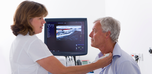
The most reliable sign of early-stage, non-enlarged LN metastasis is when perfusion - the passage of blood through the lymphatic system - shows defects, something detectable by CT scans, MRIs and ultrasound imaging devices.
Now, professor Tetsuya Kodama and his research group from Tohoku University Graduate School of Biomedical Engineering have revealed that the absence of small blood vessels in tumors may be the cause of impaired perfusion in non-enlarged, early-stage metastatic LNs.
To make their breakthrough, the research group injected cancer cells into mice bearing LNs of similar size to humans (about 10 mm). LNs injected with the cancer cells metastasized to connected LNs, where the researchers carefully tracked the cancer's progression using contrast-enhanced high-frequency ultrasound and micro-CT imaging.
"We found that tumors forming with few micro-blood vessels are associated with defective perfusion," said Kodama. "The finding could explain the low efficiency of systemic chemotherapy for metastatic LNs.
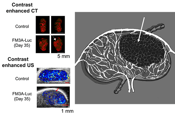
Perfusion defect in the lymph node visualized by contrast enhanced micro-computed tomography and high-frequency ultrasound (arrows).ⒸTohoku University
Their study also highlighted the importance of improving the spatial and density resolution of diagnostic imaging equipment to better improve the diagnosis and detection of LN metastasis, thereby curbing cancer-related mortality.
The research paper was published in the journal Clinical & Experimental Metastasis on October 15, 2021.
- Publication Details:
Title: Characterizing perfusion defects in metastatic lymph nodes at an early stage using high-frequency ultrasound and micro-CT imaging
Authors: Teppei Yamaki, Ariunbuyan Sukhbaatar, Radhika Mishra, Ryohei Kikuchi, Maya Sakamoto, Shiro Mori, Tetsuya Kodama
Journal: Clinical & Experimental Metastasis
DOI: 10.1007/s10585-021-10127-6
Contact:
Tetsuya Kodama,Department of Biomedical Engineering, Tohoku University Graduate School of Biomedical Engineering
Fax: +81-22-217-4818
Email: kodama
 tohoku.ac.jpWebsite: https://web.tohoku.ac.jp/kodama/