Compared with computers, the brain can perform computations with a very low net energy supply. Yet our understanding surrounding how the biological brain manages energy is still incomplete. What is known, however, is that the dilation and constriction cycles of blood vessels, or vasomotion, spontaneously occur in the brain, a process that likely contributes to enhancing the circulation of energetic nutrients and clearing wasteful materials.
Now, researchers from Tohoku University have developed a method that easily observes and monitors blood vessel dynamics in the mouse brain. This can be done either through the intact skull of a mouse, or deep into the brain using an implanted optical fiber.
The findings were detailed in the journal, eLife, on April 17, 2024.
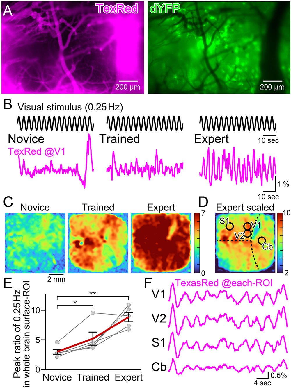
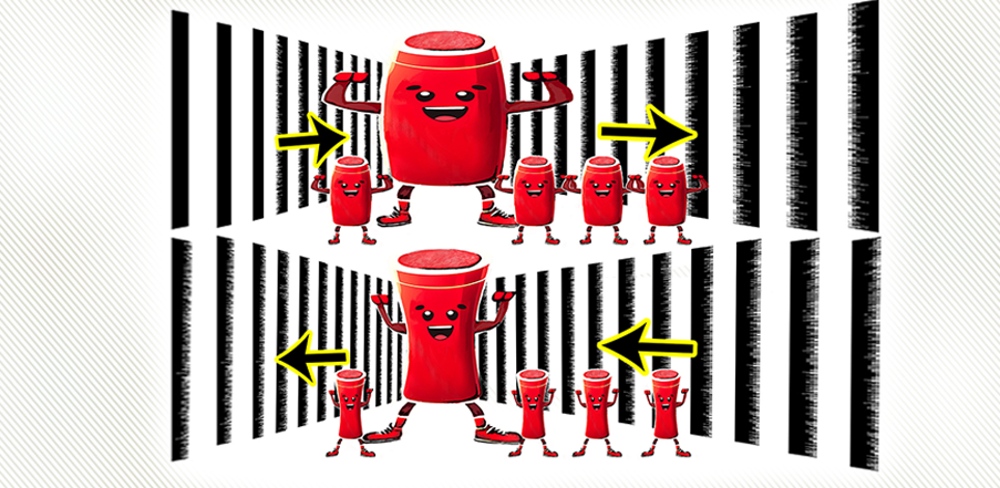
Since it has been reported that sensory stimuli can cause dilation of blood vessels or hyperemia, researchers sought to induce vasomotion via presenting mice with visual stimuli. What they discovered was when a mouse was shown a horizontally moving stripe pattern that changed direction every 2 to 3 seconds, it caused a reaction in the mouse's blood vessels that matched the pattern's speed.
Mice were presented with 15-minute visual training sessions interleaved with 1-hour resting periods for 4 times per day. With such spaced training, the amplitude of the synchronized vasomotion gradually increased. Interestingly, the visually induced vasomotion was not confined to the area of the cerebral cortex responsible for visual information processing. In other words, synchronized vasomotion spread throughout the whole brain.
"Synchronized vascular motion can be entrained with slowly oscillating visual stimuli," says Professor Ko Matsui of the Super-network Brain Physiology lab at Tohoku University, who led the research. "Such enhancement of circulation mechanisms may benefit the information processing capacity of the brain."

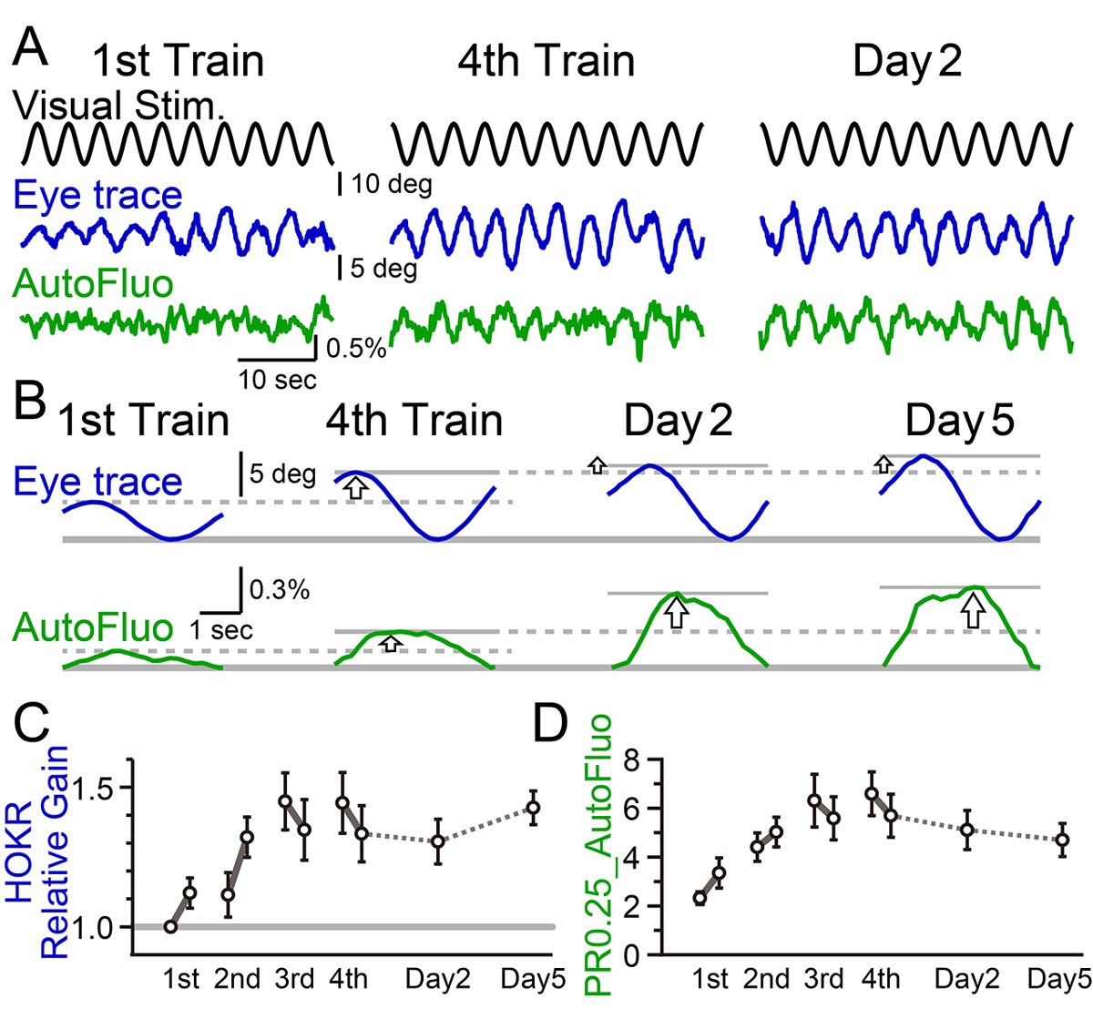
While it's long been known that changes in neuron connections support learning and memory, the plasticity of vasomotion hasn't been described before. Matsui and his colleagues found that a specific visual pattern makes the eyes move more, and this eye movement improvement depends on changes in the brain's cerebellum. The researchers also observed that blood vessel activity in the cerebellum synchronized with this optokinetic motor learning.
Lead study investigator, Daichi Sasaki, believes that synchronized vasomotion, which efficiently delivers oxygen and glucose, could improve learning abilities. He states, "Our next step is to explore the advantages of vasomotion synchronization. It might help clear waste like amyloid beta, potentially delaying or preventing dementia. Stroke recovery could also benefit from better energy supply and waste removal. Additionally, synchronized vasomotion might even enhance intelligence beyond our natural capabilities."

- Publication Details:
Title: Plastic vasomotion entrainment
Authors: Daichi Sasaki, Ken Imai, Yoko Ikoma, Ko Matsui
Journal: eLife
DOI: https://doi.org/10.7554/eLife.93721.3
Contact:
Ko Matsui,
Super-network Brain Physiology, Graduate School of Life Sciences
Email: matsui med.tohoku.ac.jp
Website: http://www.ims.med.tohoku.ac.jp/matsui/


