There are few scientific methods more elegantly simple than "just sprinkle it on top." Researchers at Tohoku University and Nagoya University developed a fluorescent probe that can quickly show synapses, the connection points between brain cells. By simply "sprinkling" this probe on top, active synapses will visibly light up so that researchers can clearly observe how living brain cells communicate - all within 10 seconds. This research gives us a new way to see when, where, and how memory is formed in the brain as well as the basis for learning.
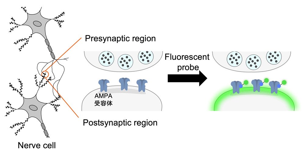
Our brain cells communicate by releasing neurotransmitters at information relay points called synapses. Some synapses are strong, meaning signals pass through easily. Other synapses are weak, therefore signals are harder to send. This strength depends on how much neurotransmitter is released, how sensitive the receptors are, and how the receiving cell responds. However, this strength is not permanently set in stone. Our brains can change via synaptic plasticity as we learn new things. For example, practicing a new language everyday could make certain synapses stronger. You also may have heard that young children have high brain plasticity in general, which is why they can pick up a second language much more quickly than an adult.
To classify how synapses behave during memory formation, the researchers looked at how AMPA receptors were delivered to synapses as their connections became stronger via a process called long-term potentiation (LTP).
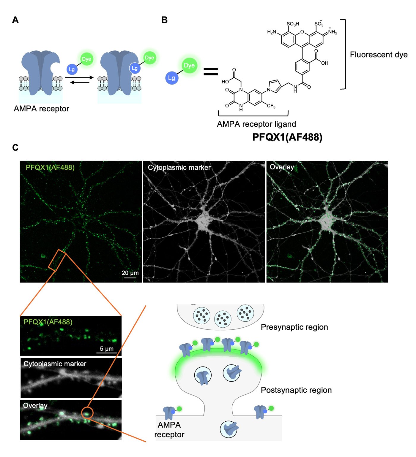
A novel fluorescent probe called PFQX1(AF488) was developed to label AMPA receptors upon living brain cells. By simply sprinkling the probe, the researchers could quickly see where AMPA receptors were on the cell surface. In real-time, they saw that during LTP, new receptors were mainly inserted from inside the cell, not moved from nearby areas. These findings help clarify the exact mechanism of AMPA receptor upregulation, especially since previous methods returned inconsistent results.
To confirm that the new receptors were coming from inside the cell via exocytosis, they applied an exocytosis-blocking drug. The increase in PFQX1 signal due to new receptor insertion disappeared, confirming exocytosis as the main way AMPA receptors reach synapses during LTP.
"Until now, it was hard to see which synapses were strong or active in living brain cells, but our new probe solves this problem," says Professor Eriko Nango (Tohoku University).
This probe provides a new, easy way to study how memories form, without requiring genetic modification or complex tools. Researchers may now be better equipped to explore memory formation, brain development, and even diseases like dementia and Alzheimer's disease in-depth.
"Our novel probe makes it easier than ever to study how brain cells communicate and how learning occurs at the molecular level. This is a powerful step forward for brain research," summarizes Professor Shigeki Kiyonaka (Nagoya University).
The findings were published in Science Advances on June 7, 2025.
- Publication Details:
Title: Rapid and reversible fluorescent probe enables repeated snapshot imaging of AMPA receptors during synaptic plasticity
Authors: Kyohei Soga, Takaaki Fujiwara, Mayu Nakagawa, Akihiro Shibata, Hansel Adriel, Kenji Yatsuzuka, Wataru Kakegawa, Michisuke Yuzaki, Itaru Hamachi, Eriko Nango, Shigeki Kiyonaka
Journal: Science Advances
DOI: 10.1126/sciadv.adt6683
Contact:
Eriko Nango
Institute of Multidisciplinary Research for Advanced Materials, Tohoku University
Email: eriko.nango.c4 tohoku.ac.jp
Website: https://www2.tagen.tohoku.ac.jp/lab/nango/html/en/index.html
Shigeki Kiyonaka
Graduate School of Engineering, Nagoya University
Email: kiyonakachembio.nagoya-u.ac.jp
Website: https://www.chembio.nagoya-u.ac.jp/labhp/life1/index.html