Anxiety is often attributed to an unconscious assessment of the environment and detection of potential danger. Whilst moderate anxiety is therefore advantageous for survival, excessive anxiety can lead to psychiatric disorders.
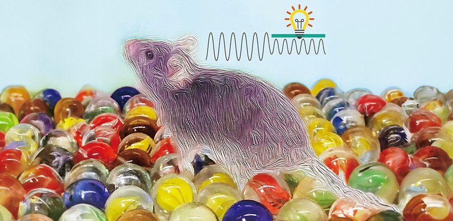
Now, researchers at Tohoku University have shed light on the intricate interactions between neurons and astrocytes within the habenula, a region of the brain associated with emotional processing. By subjecting mice to a scenario involving a floor scattered with marbles, the researchers observed behavioral responses indicative of anxiety.
The findings were detailed in the journal, Neuroscience Research, on February 10, 2024.
The habenula are a pair of small nuclei located above the thalamus. It is one of the few brain regions that controls both dopaminergic and serotonergic systems. As these neuromodulators play essential roles in a wide range of motivational and cognitive functions, habenula neuronal circuits are potentially relevant to controlling anxiety.
"Anxiety may appear to be an irrational emotion having only negative impacts on our life," says Professor Ko Matsui of the Super-network Brain Physiology lab at Tohoku University, who led the research. "However, well-tuned anxiety is a guide provided by our unconsciousness which allows us to navigate the hidden dangers. Such tuning may be accomplished by the actions of the habenula."
Mice perceive smooth glass marbles as potentially harmful objects due to their unfamiliarity. Mice tend to bury marbles in saw dust bedding to keep these uncomfortable objects out of sight. Here, the researchers created a chamber filled with marbles to create an inescapable, maximum anxiety environment.
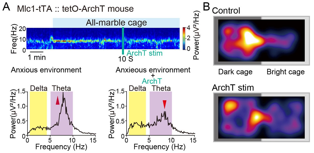
They noticed increased neuronal activity in the theta band (5 to 10 Hz) frequency, an increase in local brain blood volume, and acidification occurring in the astrocytes of the habenula when the mice were placed in the all-marble cage. When the habenular astrocytes were artificially alkalized to counter the acidification, the theta band neuronal activity diminished. When the mice were allowed to choose between the brightly lit all-marble cage and a dark and comfortable cage, the mice naturally chose to stay in the dark cage. However, when the habenular astrocytes were optogenetically alkalized, the mice ventured more into the bright cage.
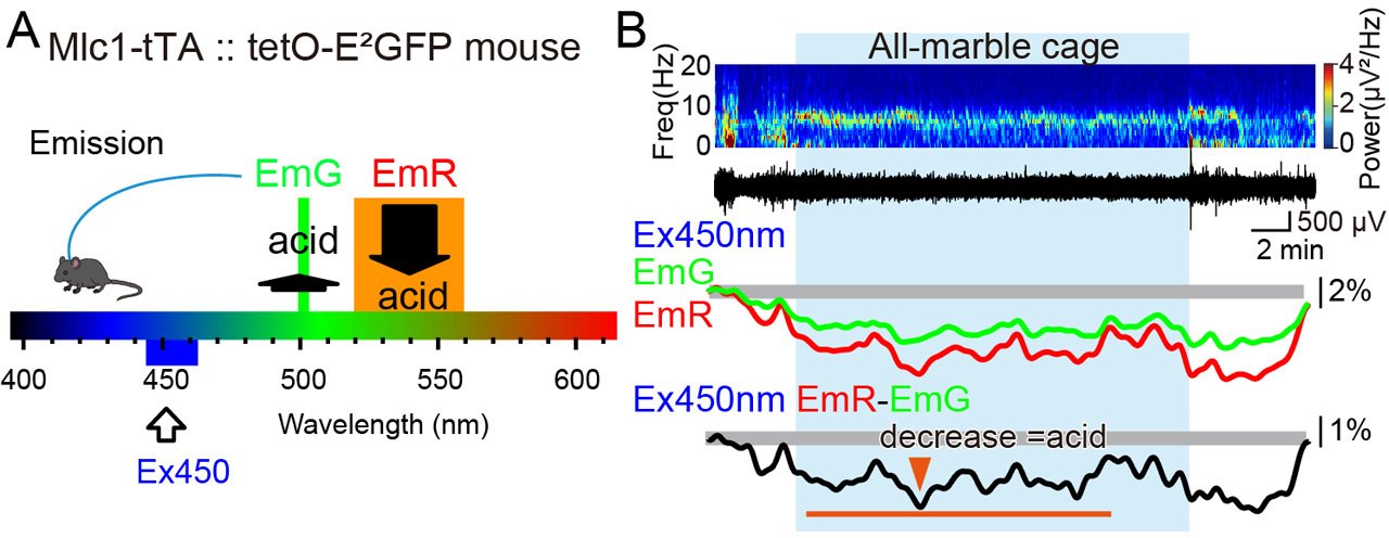
Astrocytes are non-neuronal cells that occupy approximately half of the brain. They have been shown to control the local ionic and metabotropic environment in the brain. Astrocytes also release transmitters that can affect neuronal activity in the vicinity. The results of this study suggest that the theta band habenular neuronal activity is regulated by the activity of astrocytes. Thus, habenular astrocytes were considered to play a role in regulating anxiety.
Lead study investigator, Wanqin Tan, says that future treatment of anxiety disorders may be realized by developing a therapeutic strategy that adjusts astrocyte activity in the habenula. "Habenular astrocytes tune the 'marble blues.' Based on this, we expect that methods to cope with anxiety could be developed."
- Publication Details:
Title: Anxiety control by astrocytes in the lateral habenula
Authors: Wanqin Tan, Yoko Ikoma, Yusuke Takahashi, Ayumu Konno, Hirokazu Hirai, Hajime Hirase, Ko Matsui
Journal: Neuroscience Research
DOI: https://doi.org/10.1016/j.neures.2024.01.006
Contact:
Ko Matsui
Super-network Brain Physiology, Graduate School of Life Sciences,
Tohoku University
Email: matsui med.tohoku.ac.jp
Website: http://www.ims.med.tohoku.ac.jp/matsui/