Researchers at Tohoku University in Japan have collaborated with others to develop a simple way to create and functionalize virus-like polymer particles that have various nanostructures. The collaboration includes researchers from Michigan University in the USA and Karlsruhe Institute of Technology (KIT) in Germany.
The geometrical control of enzymes, antibodies and other proteins over polymer particles is essential for realizing cascade reactions observed in a living body; highly sensitive immunoassay systems; and highly efficient drug delivery systems. Given enzymatic reactions occur step-by-step along aligned enzymes, the formations of such enzyme arrays are crucial for realizing the particles.
In immunoassay systems and drug delivery systems using polymer particles, the density and alignment of antibodies on the particles are very important factors in achieving high sensitivities. A virus is an ideal particle given its nanostructures and geometrically controlled functional groups. However, structural control and selective chemical modification of synthetic polymer particles have until now been inaccessible due to their complicated synthetic routes.
Diblock copolymers form nanoscale phase-separated structures, and macromolecular compositions can control structures and periodicities. In the present study, Guillaume Delaittre and co-workers at KIT have succeeded in synthesizing hydrophobic diblock copolymers that have functionalized blocks. Divya Varadharajan at KIT and Hiroshi Yabu at Tohoku University have converted those diblock copolymers to nanostructured particles by using a nanoprecipitation method they developed.
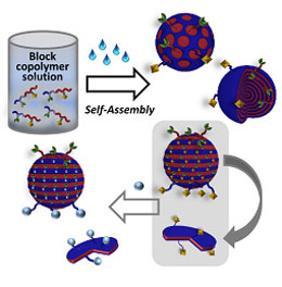
Schematic illustration of the work.
Credit: John Wiley & Sons, Inc.
Changing the preparation conditions, sizes and morphologies of particles led to core-shell, stacked lamellae, and other morphologies being found. The stacked lamellae structure, in which both polymer phases are exposed to the particle surfaces, was chosen for selective chemical modification.
To visualize the site-selective chemical modification of particles, fluorescent dyes were fixed on the one polymer phase. Joerg Lahann at Michigan University identified this chemical modification by observing ring-shaped fluorescence from nanodiscs originating from disassembling stacked lamellae. Lahann used stimulated emission depletion (STED) microscopy in his work, which is one of the super-resolving microscopy methods.
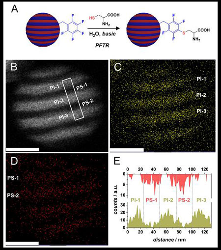
(A) Schematic representation of functionalized block of the striped nanoparticles with cysteine. (B) Dark field STEM image of nanoparticles: the bright parts (PI-1-3) represent the polyisoprene (PI) segment; the dark regions represent the polystyrene (PS) segment. (C) and (D) Elemental analysis of non-functionalized PI region and functionalized PS region, respectively. (E) Grey value pixel map obtained by integrating the area in the marked region in (B) showing PS segments PS-1-2 and PI segments PI-1-3 showing alternate stacks of S (red) and Os (green) that represent PS and PI segments respectively. All scale bars represent 50 nm.
Credit: John Wiley & Sons, Inc.
- Publication Details:
Title: Surface-Reactive Patchy Nanoparticles and Nanodiscs Prepared by Tandem Nanoprecipitation and Internal Phase Separation
Authors: Divya Varadharajan, Hatice Turgut, Joerg Lahann, Hiroshi Yabu*, and Guillamume Delaittre*
Journal: Advanced Functional Materials
DOI:10.1002/adfm.201800846
Contact:
Hiroshi YabuAdvanced Institute for Materials Research (AIMR)Tohoku University
Email: hiroshi.yabu.d5
 tohoku.ac.jp
Website: https://www.wpi-aimr.tohoku.ac.jp/yabu_labo/index/Welcome.html